The Last Step of Every Breath
Air is moved by pressure. Blood is moved by the heart. But the final micrometres into a living cell have no pump behind them — only random motion and a difference in concentration.
A drop of dye
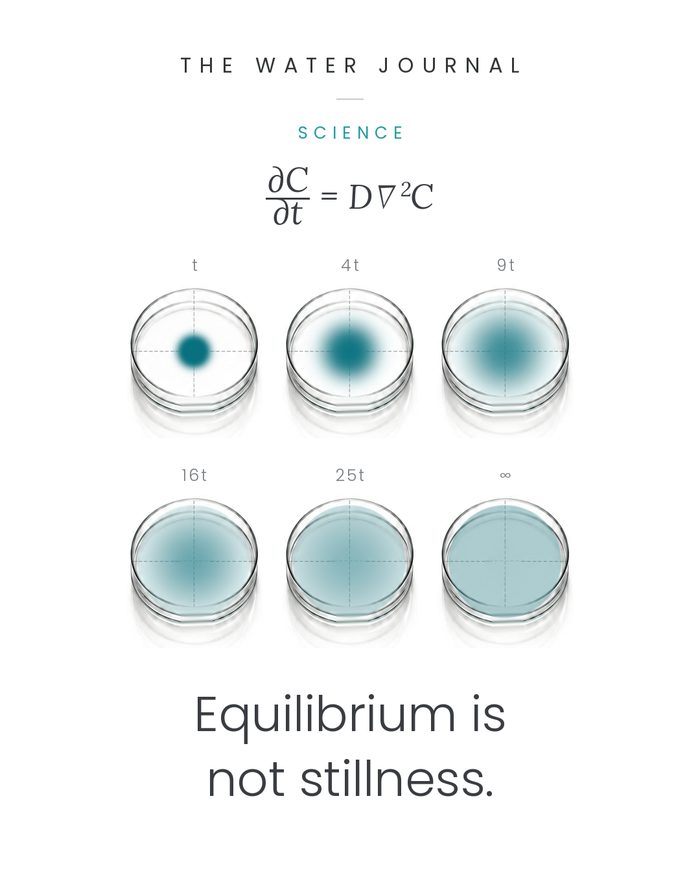
Put a single drop of dye into a dish of still water and photograph it every so often. The dye spreads. Nothing stirs it. No current carries it. Left alone, it fills the dish and then stops changing.
Now measure the spread rather than watch it. It does not keep pace with the clock. To double the distance the dye has travelled you must wait four times as long. To triple it, nine times. The dishes on the cover of this issue are labelled t, 4t, 9t, 16t, 25t for exactly that reason: they illustrate free-diffusion scaling, where — before the walls of the container begin to matter — equal steps in characteristic diffusion length fall at those times. Time sits on one axis and distance on another, and the two are not the same shape. The dishes on the cover are labelled t, 4t, 9t, 16t, 25t for exactly that reason: those are the moments at which the characteristic diffusion length has increased by equal steps. Time sits on one axis and distance on another, and the two are not the same shape.
That single mismatch — distance advancing as the square root of time — helps dictate the anatomy of every animal large enough to see.
Randomness makes a direction
At the molecular scale, stillness is an illusion. Molecules in a fluid move and collide continuously because of thermal motion, and each individual path is irregular. A given molecule may drift toward a thinner region, then immediately drift back.
Diffusion does not require any molecule to know where it is going. It requires only an imbalance in how many are available to move. Picture a boundary with many dye molecules on one side and few on the other. Molecules cross in both directions at random — but more of them start on the crowded side, so more cross away from it than return. The individual motion stays random; the sum of it does not. What emerges is a net movement from higher concentration toward lower: orderly, reliable, and produced entirely by disorder.
Einstein's 1905 treatment of Brownian motion put this on a statistical footing, connecting the erratic path of a single particle to the smooth spreading of a population of them.
Molecules do not seek equilibrium. Equilibrium is what probability leaves behind.
Fick's two laws
Half a century before Einstein, the German physiologist Adolf Fick wrote the behaviour down. His 1855 paper gives the relation still in use today.
J is the diffusive flux, D the diffusion coefficient of that substance in that medium, and ∇C the concentration gradient. Almost the whole idea sits in the minus sign: net movement runs down the gradient, from more to less.
His second law describes not the flow at one instant but how the entire pattern changes over time.
A concentrated spot flattens. Its centre thins, its edges reach outward, and in a closed dish the concentration eventually becomes even throughout.
At that point something worth noticing happens. The molecules do not stop. They keep crossing in both directions — but at equilibrium the opposing fluxes are equal, and the net flux is zero. What ends is not the motion. What ends is the gradient.
Why distance is the tyrant
Fick's second law carries a scaling law inside it. The characteristic time for diffusion rises roughly with the square of the distance to be covered. Double the distance, wait about four times as long. Increase it tenfold, and the timescale grows about a hundredfold.
Across a micrometre, diffusion is extraordinarily fast. Across a metre, it is useless.
Your body does not attempt to diffuse oxygen from the lungs to the toes. It moves air by pressure and blood by pumping — bulk flow, vastly more effective across macroscopic distances than diffusion — and it uses that flow to carry oxygen to within a few micrometres of where it is needed. Only then is diffusion handed the job.
The cardiovascular system is not an alternative to diffusion. It is the infrastructure that keeps diffusion's distances short enough to be survivable.
Water is the medium, and the limit
Much of this final journey occurs through water-rich biological media: blood plasma, interstitial fluid, the cytoplasm of the cell — and across the membranes and barriers that separate them.
Water does not tell oxygen where to go. The partial-pressure gradient does that. But the local medium helps set how quickly oxygen can move, and water is not generous. Oxygen diffuses through air roughly ten thousand times faster than through water. The same molecule, the same random motion, the same physics — and four orders of magnitude of difference, made by nothing but the medium.
This is the constraint the lung is built against. If the barrier between alveolar air and capillary blood were as thick as ordinary tissue, gas exchange would be far too slow to support a mammal. So it is not. In a classic 1978 morphometric study of healthy human lungs, the harmonic mean thickness of that barrier was about 0.62 micrometres, folded out across roughly 143 square metres of alveolar surface. Thin enough to cross quickly; wide enough to cross a great deal at once.
Life performs this final step through water-rich tissue. That medium sustains the chemistry of the cell — and imposes a severe diffusion penalty.
Given a gradient, enough random steps become a direction.
The last micrometre
Follow one breath to the end. Air arrives in the alveoli. Oxygen crosses that sub-micrometre barrier into capillary blood, where haemoglobin binds it and multiplies what the blood can carry.
Even here diffusion is not finished: oxygen still has to move through the interior of the red cell itself. Work published in 2020, imaging oxygen exchange in single human red cells, found that diffusion through their haemoglobin-rich cytoplasm is itself a dominant resistance to gas transport. A cell built almost entirely for carrying oxygen still cannot outrun the physics of moving it.
Downstream, in the microcirculation, metabolism keeps consuming oxygen and so holds the oxygen partial pressure in the surrounding tissue below the partial pressure inside the vessel. Oxygen leaves the capillary and spreads through the tissue. Measurements in rat mesentery have reported intraluminal and perivascular pO₂ gradients around arterioles. Their magnitude, however, has been subject to methodological debate: the phosphorescence technique used to make them can consume oxygen as it measures, and so exaggerate an apparent drop.
The circulation has now done everything it can do. There is no artery for the last stretch and no heartbeat that reaches it. The last stretch is molecular, and it is crossed by molecules that have no destination in mind.
The same physical principle runs from a drop of dye in a dish to the exchange surfaces that keep a human being alive.
Sources and notes
The physics. Adolf Fick, "Ueber Diffusion," Annalen der Physik, vol. 170, no. 1 (1855), pp. 59–86 — first statement of the flux law. Albert Einstein, "Über die von der molekularkinetischen Theorie der Wärme geforderte Bewegung von in ruhenden Flüssigkeiten suspendierten Teilchen," Annalen der Physik, vol. 322, no. 8 (1905), pp. 549–560 — statistical basis for diffusion length growing as the square root of time.
The measurements. P. Gehr, M. Bachofen and E. R. Weibel, "The normal human lung: ultrastructure and morphometric estimation of diffusion capacity," Respiration Physiology, vol. 32 (1978), pp. 121–140 — air–blood barrier harmonic mean thickness 0.62 ± 0.04 µm; alveolar surface area 143 ± 12 m². G. A. Somerville and R. A. Proctor, "Cultivation conditions and the diffusion of oxygen into culture media," BMC Microbiology, vol. 13 (2013), art. 9 — oxygen diffusion coefficient 2.1 × 10⁻⁵ cm²/s in water at 25 °C against approximately 0.2 cm²/s in air.
The physiology. S. L. Richardson et al., "Single-cell O₂ exchange imaging shows that cytoplasmic diffusion is a dominant barrier to efficient gas transport in red blood cells," PNAS, vol. 117, no. 18 (2020), pp. 10067–10078. A. G. Tsai et al., "Microvascular and tissue oxygen gradients in the rat mesentery," PNAS, vol. 95, no. 12 (1998), pp. 6590–6595 — intraluminal and perivascular pO₂ measured by phosphorescence quenching in living tissue. A. S. Golub and R. N. Pittman, "PO₂ measurements in the microcirculation using phosphorescence quenching microscopy at high magnification," American Journal of Physiology — Heart and Circulatory Physiology, vol. 294, no. 6 (2008), pp. H2905–H2916 — shows that oxygen photoconsumption by the measuring method can itself produce apparent transmural and longitudinal pO₂ gradients in arterioles.
On the evidence. Tsai et al. 1998 is an animal model (rat mesentery), and the magnitude of the gradients it reports is disputed: Golub and Pittman 2008 show that the measurement technique can consume oxygen and inflate an apparent transmural drop. Cited here for the existence of a gradient from vessel into tissue, not for its size, and not as human values. Richardson et al. 2020 is a single imaging study, cited for the resistance it identifies inside the red cell rather than as a settled quantity. Fick 1855 and Einstein 1905 are dated by publication, not by discovery.